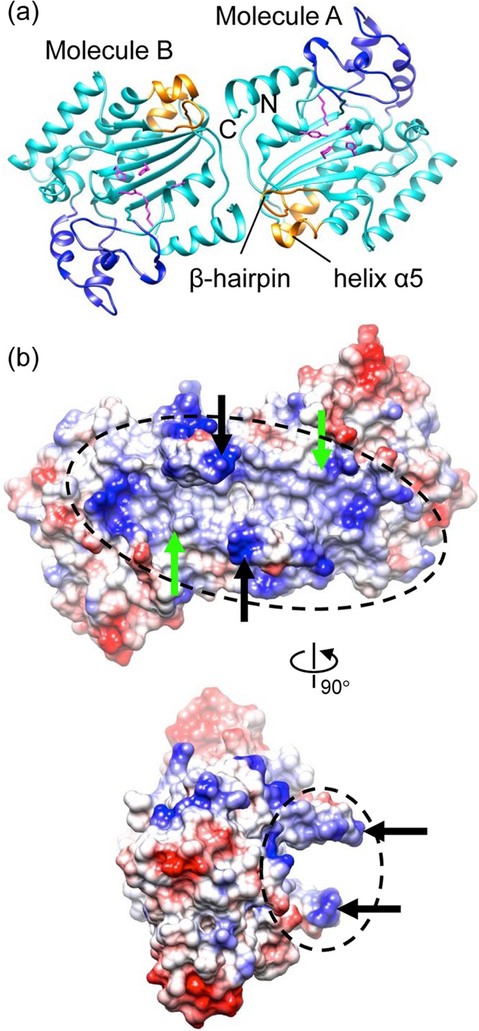
Fig 4.

Crystallographic dimer. (a) Ribbon diagram of the dimer (cyan) with the subdomain, the putative Ori-binding site, and the nuclease active site residues shown in blue, gold, and magenta, respectively. (b) Electrostatic potential molecular surface of the dimer showing an extended positively charged area (dashed ellipsoids) spanning two nuclease active sites (active Tyr indicated with green arrows) and the putative Ori recognition sites (β-hairpin indicated with black arrows). The bottom view is 90° about the vertical axis from the top view.
