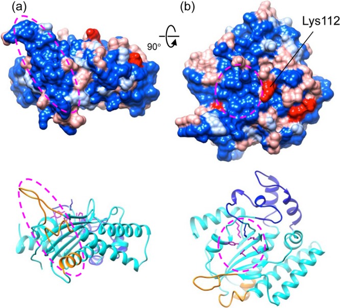
Fig 5.

Mapping the variable amino acid residues onto the HBoV-NS1N structure. The amino acid sequences of NS1 from the four HBoV species were aligned, and the residues that are constant among four, three, and two sequences and different for all four sequences are colored blue, light blue, light red, and red, respectively. The putative Ori recognition site and the nuclease active site are indicated with an ellipsoid in panels a and b, respectively. One of the highly variable residues, Lys112, which is next to the active site, is indicated. The view in panel a is 90° about the horizontal axis from that in panel a. In panels a and b, ribbon diagrams of the structure in the same view colored in the same scheme as in Fig. 4a are shown at the bottom.
