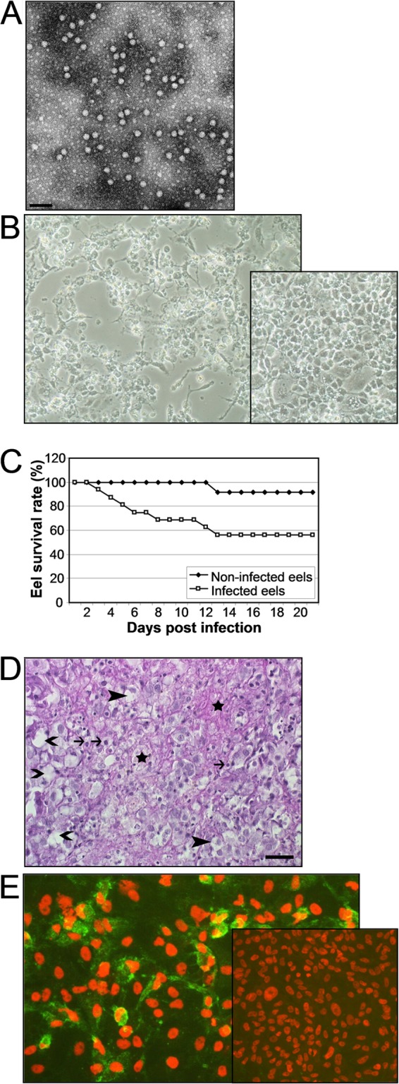
Fig 1.

Characterization of eel picornavirus. (A) Electron micrograph of EPV-1 from supernatant of infected EK-1 cells. Bar, 100 nm. (B) Cytopathic effect of EK-1 cells induced by EPV-1 after 3 days p.i. The inset shows uninfected cells. (C) Survival rates of infected and control eels. Glass eels (n = 16) were infected with EPV-1 by bathing for 1 h in virus-containing water. A control group of 12 glass eels was mock infected. The eels were kept in aquaria for 21 days. One glass eel of the control group died after an injury. (D) Histopathological changes in the liver of an experimentally infected glass eel. Hepatocytes are irregularly vacuolated (closed arrowheads). Multiple cell membranes are disrupted (open arrowheads). There are karyopyknosis (arrows) and small areas of necrosis and fibrin exudation (stars). Bar = 25 μm. Hematoxylin-eosin stain. (E) Detection of EPV-1 by indirect immunofluorescence using the rabbit antiserum T51 (in-house production) and goat anti-rabbit IgG–FITC fluorescein isothiocyanate (Sigma), with counterstaining of nuclei with propidium iodide. The inset shows uninfected cells.
