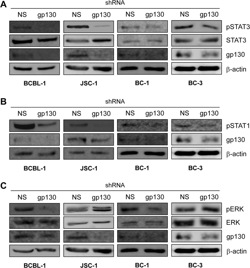
Fig 2.
Contribution of gp130 signaling to levels of active STATs and ERKs in PEL cells. Phosphotyrosine-specific antibodies were used in immunoblotting to detect and determine the levels of phosphorylated (active), gp130-targeted STAT3 (A), STAT1 (B), and ERKs 1 and 2 (C) (pSTAT3, pSTAT1, and pERK) present in lysates of PEL cells depleted of gp130 relative to the levels of these proteins in NS shRNA-transduced controls. Antibodies detecting total (phosphorylated and unphosphorylated) STAT3 and ERK1/2 and β-actin were used to verify equivalent protein loading on SDS-PAGE gels and transfer to blotting membranes. For each set of lysates (used for single- or multiple-phosphoprotein detection), effective depletion of endogenous gp130 in the lentiviral shRNA-infected PEL cell cultures was verified by immunoblotting for gp130. Monitoring of growth of NS shRNA- versus gp130 shRNA-transduced cultures for BCBL-1, JSC-1, and BC-1 cells over 3 days (as described in the legend to Fig. 1) confirmed the growth-inhibitory effects of gp130 depletion (data not shown); cells were harvested on day 3 for Western analyses. For BC-3 cultures, all cells were harvested 48 h after lentiviral infection (day 0) to ensure sufficient material for immunoblotting.

