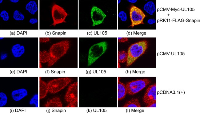
Fig 5.

Colocalization of UL105 and Snapin expressed in human cells. Cells were cotransfected with constructs pCMV-Myc-UL105 and pRK11-FLAG-Snapin (a to d), pCMV-UL105 alone (e to h), or pCDNA3.1(+) alone (i to l), fixed at 48 h posttransfection, stained with DAPI (a, e, and i), anti-FLAG (b), anti-Myc (c), anti-Snapin (f and j), and anti-UL105 (g and k) antibodies, and visualized using a confocal microscope (Olympus FV1000). The images of Snapin (b, f and j), UL105 (c, g and k), and the nuclei stained with DAPI (a, e, and i) were used to generate the composite images (d, h, and l). The images show different levels of magnification.
