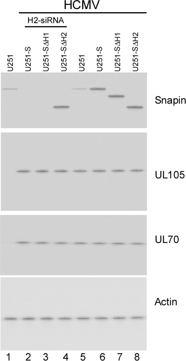
Fig 6.

Expression of Snapin and its mutants in cells. Western blot analysis was used to determine the levels of Snapin, UL70, and UL105 in the parental U251 cells (U251) (lanes 1 and 5), cells that expressed full-length Snapin (U251-S; lanes 2 and 6), and Snapin mutants SΔH1(37-65aa) (U251-SΔH1; lanes 3 and 7) and SΔH2(81-126aa) (U251-SΔH2; lanes 4 and 8). Cells were either transfected with anti-Snapin H2-siRNA molecules (lanes 2 to 4) or not transfected with any siRNAs (lanes 1 and 5 to 8) and were either mock infected (lane 1) or infected with HCMV at an MOI of 1 after 48 h of siRNA incubation (lanes 2 to 8). The expression of cellular actin was used as the internal loading control.
