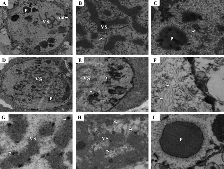
Fig 5.
Electron microscopy of Sf9 cells transfected with vAc83KO or vAc83:HA. (A to C) Cells transfected with vAc83:HA. (A) Portion of a cell showing the virogenic stroma (VS), polyhedra (P), and nuclear membrane (nm) (arrow). (B) Rod-shaped nucleocapsids (arrow) within the electron-dense edges of the VS. (C) Multiply enveloped mature ODVs (arrow) and polyhedra with ODVs embedded. (D to I) Cells transfected with vAc83KO. (D) VS. (E) Portion of the cell region showing microvesicles (arrows) in the nucleus. (F) Cluster of elongated, electron-lucent tubular structures that localized near the inner nuclear membrane (arrows). (G) VS devoid of nucleocapsids and exhibiting electron-dense bodies (arrows). (H) Nucleus with aberrant tubular structures (arrows) within the VS. (I) Polyhedra devoid of embedded virions.

