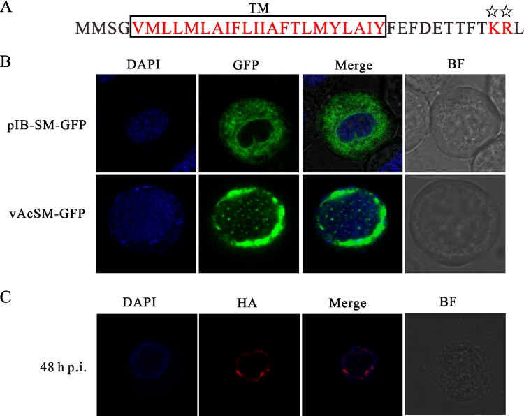
Fig 9.
Ac83 contains a functional inner nuclear membrane sorting motif and is localized to the nuclear periphery during infection. (A) Schematic representation of two characteristics of the INM-SM of Ac83. The highly hydrophobic TM is boxed and shown in red. The conserved positively charged amino acids are also shown in red and are marked with stars. (B and C) Confocal microscopy. (B) Sf9 cells were transfected with pIB-SM-GFP (top) or were infected with vAcSM-GFP (bottom) at an MOI of 5 TCID50s/cell. At 24 h p.t. or 48 h p.i., the cells were prepared for monitoring of the localization profile of GFP (green). (C) Sf9 cells were infected with vAc83:HA at an MOI of 5 TCID50s/cell. At 48 h p.i., the cells were fixed, probed with a mouse monoclonal anti-HA antibody to detect HA-tagged Ac83, and visualized using Alexa Fluor 647-conjugated goat anti-mouse IgG (red). The cells were stained with DAPI to directly visualize nuclear DNA (blue). BF, bright-field microscopy.

