Fig 2.
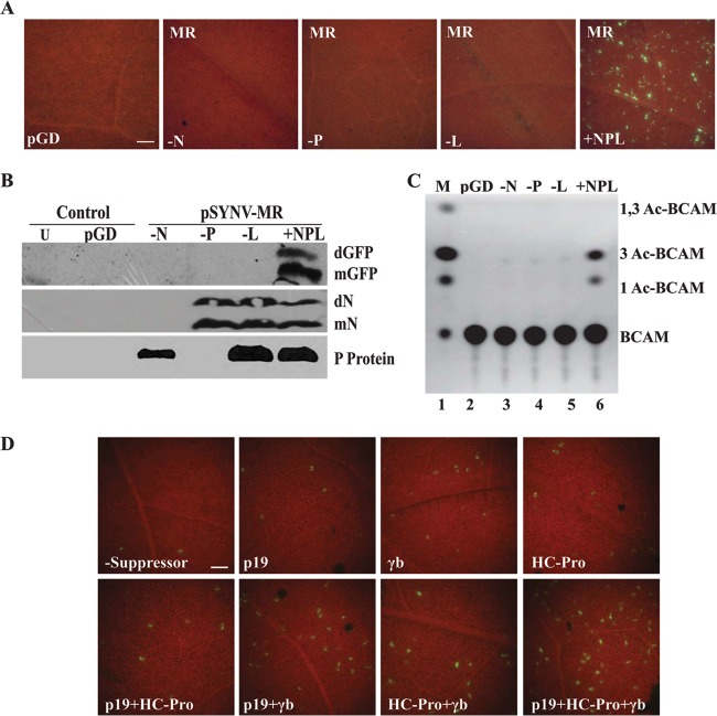
SYNV core protein requirements and gene silencing suppressor effects on GFP expression. (A) GFP foci in N. benthamiana leaves at 6 dpi with bacteria harboring the pSYNV-MReGFP-DsRed reporter cassette. The pGD panel shows a control pGD plasmid not encoding GFP. The −N, −P, and −L panels illustrate tissue infiltrated with pSYNV-MReGFP-DsRed and SYNV core protein mixtures lacking the N, P, or L plasmid, respectively. The +NPL panel shows fluorescence observed as single-cell foci scattered throughout the infiltrated regions of N. benthamiana leaves agroinfiltrated with mixtures containing the pSYNV-MReGFP-DsRed reporter cassette and the SYNV N, P and L plasmids. All experiments were conducted in the presence of p19 and γb gene silencing suppressors. The bar in the pGD panel represents 100 μm, and the remaining panels are the same magnification. (B) Western blot analysis showing GFP and SYNV N and P protein expression. Protein extracts recovered from leaf tissue were detected by immunoblotting with primary mouse GFP antibodies and secondary goat anti-mouse–horseradish peroxidase (HRP) antibodies or with rabbit antibodies elicited against the SYNV N and P proteins and secondary goat anti-rabbit–HRP antibodies. Lanes indicate uninfiltrated tissue (U), pGD-infiltrated tissue (pGD), and tissue infiltrated with pSYNV-MReGFP-DsRed lacking N (−N), P (−P), or L (−L), or pSYNV-MReGFP-DsRed containing N, P, and L (+NPL). mN, N protein monomer; dN, N protein dimer; other designations are as in Fig. 1. (C) Requirement of the N, P, and L proteins for CAT expression from SYNV plasmids in N. benthamiana tissue. Leaves were infiltrated with mixtures of bacteria containing various combinations of the N, P, and L plasmids for core protein expression, pSYNV-MRrsGFP-CAT, and the TBSV p19 and BSMV γb gene silencing suppressors. Designations along the side of the TLC plate are as in Fig. 1. (D) Effects of viral gene silencing suppressors on GFP expression from the pSYNV-MReGFP-DsRed reporter. N. benthamiana leaves were agroinfiltrated with plasmids for expression of the N, P, and L proteins and pSYNV-MReGFP-DsRed in the absence or presence of various combinations of the gene silencing suppressors TBSV p19, BSMV γb, and TEV HC-Pro. The numbers of GFP foci were substantially lower in the absence of gene silencing suppressors (−Suppressor) than in their presence, and a synergistic increase in the numbers of fluorescent cells occurred upon addition of two or more suppressors. The bar in the −Suppressor panel represents 100 μm, and the remaining panels are the same magnification.

