Fig 2.

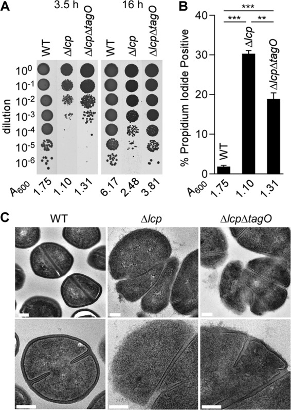
Deletion of tagO does not improve growth of the Δlcp mutant. (A) Staphylococcal viability was examined by plating serial dilutions (0- to 6-fold) of culture aliquots of the wild type and the Δlcp and Δlcp ΔtagO mutants that had been grown for 3.5 and 16 h. Images of the agar plates are shown along with the A600 values of the cultures at the time of plating. (B) Membrane integrity of staphylococci was assessed with propidium iodide staining. Culture aliquots of the wild type and the Δlcp and Δlcp ΔtagO mutants that had been grown for 3.5 h were fixed with paraformaldehyde and stained with SYTO 9 (total cells) and propidium iodide. SYTO 9-positive cells were analyzed for propidium iodide staining using flow cytometry. Combined data from two independent experiments with triplicate analyses of 20,000 cells per sample are presented. Statistical significance was determined using the Student t test. **, P < 0.01; ***, P < 0.001. (C) Transmission electron micrographs of wild-type, Δlcp, and Δlcp ΔtagO bacteria. Cells were cultured for 3.5 h, as described for panel A. Bars, 200 nm.
