
Fig 5.
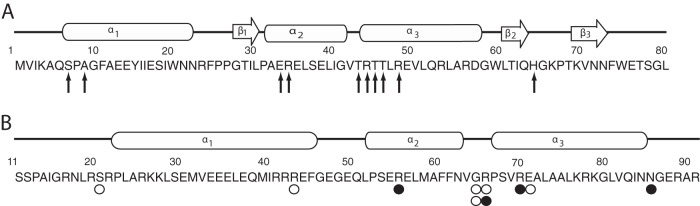
Functional comparison of FadR and NanR N-terminal DNA binding domains. Amino acid residues are numbered from the N terminus of FadR (A) or NanR (B) and are shown below the known or predicted regions of α-helical, β-helical, or random helical structures. Arrows indicate residues known to contact operator DNA nucleotides. Open circles indicate residues found altered in transdominant mutants isolated after random mutagenesis with hydroxylamine (H), while closed circles represent alterations resulting from site-directed mutagenesis (S) as described in the text.