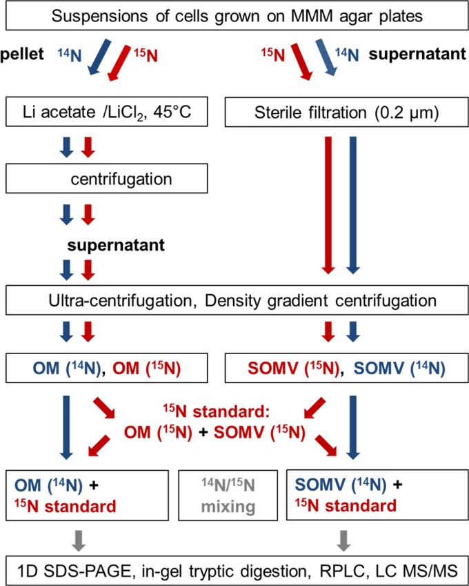
Fig 1.
Work flow of proteome analysis. OMs were obtained with lithium chloride-lithium acetate at 45°C. Density gradient centrifugation of the SOMV and OM fractions was performed to reduce cytosolic protein contamination. A 15N-labeled standard was mixed with the unlabeled samples. Proteins were digested in gel and then subjected to reversed-phase LC (RPLC). LC-MS/MS was performed with high resolution and high mass accuracy. 1D, one dimensional.

