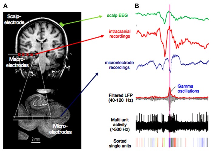
FIGURE 3.
Multiscale recordings. (A) Scalp-electrode (green), clinical multi-contact macro-electrode (red), and micro-electrode emerging from the tip of the macro-electrode (resolution: volume <1 mm3 on a millisecond scale). Such recording setups are used for presurgical evaluation in epilepsy. (B) Signal from scalp-, macro-, and micro electrode in green, red, and blue, respectively. Lower three traces show micro-electrode recordings filtered in the gamma band, with applied high-pass filter above 500 Hz and sorted spikes for different neurons. Note the high-frequent activity present in the micro-electrode recording, which is not visible in the signal from macro- or scalp-electrodes.

