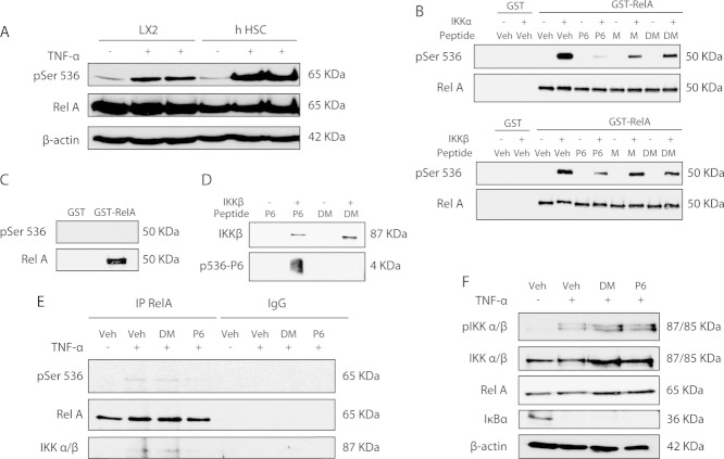
Fig 1.
P6 is a competing peptide, not an IKK inhibitor. Western blotting showing increased RelA-P-Ser536 in LX2 and hHM cultured in 0.5% FBS after stimulation with 100 ng/mL of TNF-α (A). P6 reduces GST-RelA-P-Ser536 by IKK-α and-β (B) in an in vitro kinase assay. Kinase assay input control (C). Western blotting of IKK-β kinase assay supernatant shows that P6 is phosphorylated at Ser536 by IKK-β (D). Iimmunoprecipitation for RelA in passage 4 hHM ± 100 µM of peptides for 1 hour, then stimulated with 100 ng/mL of TNF-α for 10 minutes (E). Input control for RelA immunoprecipitation (F). Gels are representative of at least three separate experiments.

