Fig 4.
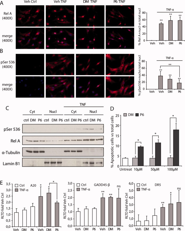
RelA nuclear translocation is RelA-P-Ser536 independent in hepatic myofibroblasts. Epifluorescence images at ×400 magnification and quantification of nuclear translocation in hHMs of RelA (red) (A) or RelA-P-Ser536 (red) (B) and 4′,6-diamidino-phenylindole (blue) in P4 hHMs in 0.5% serum-stimulated ± 100 µM of peptides for 1 hour and 50 ng/mL of TNF-α for 10 minutes. Western blotting of cytoplasmic (Cyt) and nuclear (Nucl) extracts isolated from LX2-cells–stimulated ± 100 µM of peptides and TNF-α (C). Acridine orange apoptosis assay in hHMs ± 10-100 µM of peptides 24 hours (D). A20, GADD45-β, and DR5 gene expression in LX2 cells in 0.5% serum-stimulated ± 100 µM of peptides overnight and 50 ng/mL of TNF-α for 5 hours (E). Data are expressed as mean ± standard deviation, compared to control group, and is representative of at least three separate experiments.

