Fig 6.

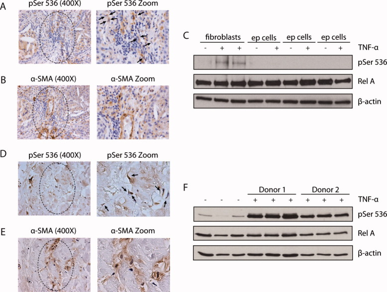
RelA-P-Ser536 is a feature of human lung and skin fibroblasts and is regulated by TNF-α. Representative photomicrographs of RelA-P-Ser536 (A) and α-SMA (B) staining in human IPF slides; black arrows denote RelA-P-Ser536-stained cells. Western blotting showing RelA-P-Ser536 and total RelA in passage 3 human lung fibroblasts or epithelial cells cultured with 0.5% FBS and stimulated with 50 ng/mL of TNF-α for 10 minutes (C). Representative photomicrographs of RelA-P-Ser536 (D) and α-SMA (E) staining of human consecutive slides of sclerodema; black arrows denote RelA-P-Ser536-stained cells. Western blotting showing RelA-P-Ser536 and total RelA human healthy skin fibroblasts cultured with 0.5% FBS and stimulated with 50 ng/mL of TNF-α for 10 minutes (F). Data represent at least two separate cell lines and 3 IPF and scleroderma patients.
