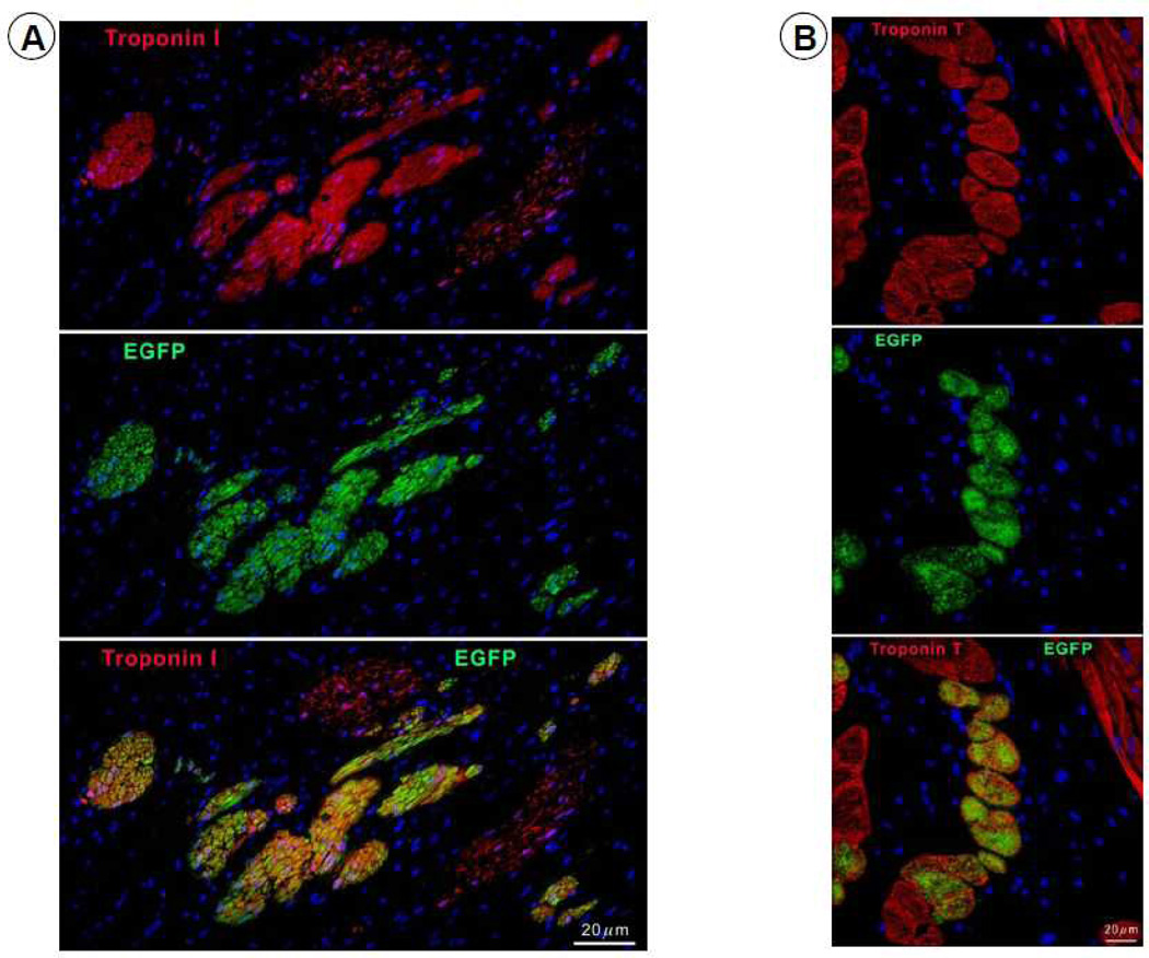
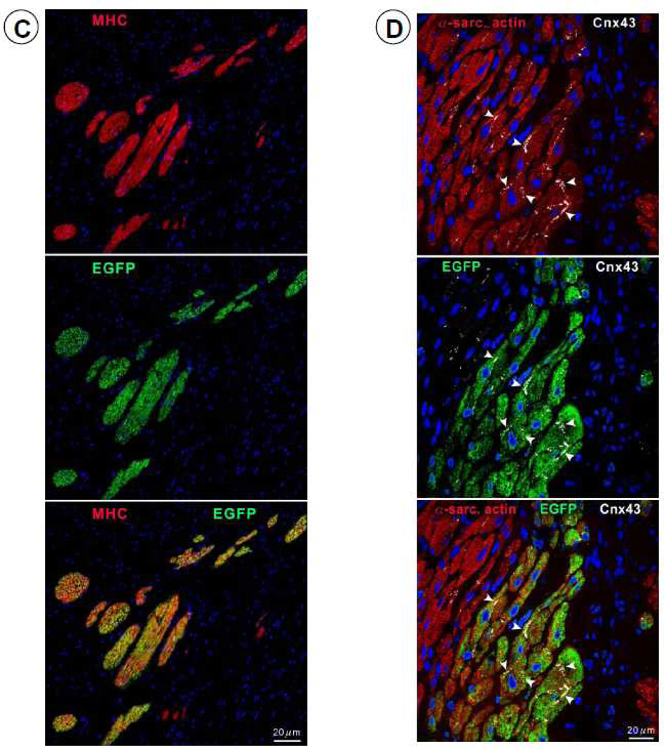
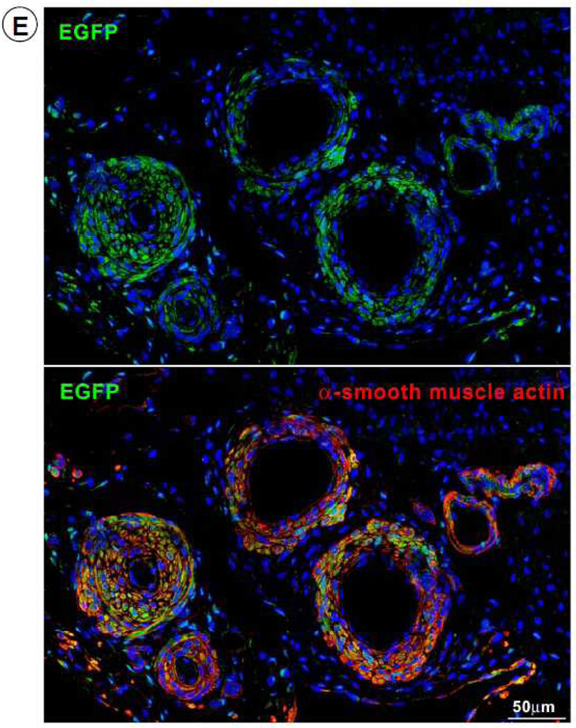
Figure 8.
Expression of cardiac-specific TnI, TnT, MHC, Cnx43, and vascular smooth muscle protein (α-smooth muscle actin) in EGFP-positive cells. Representative confocal microscopic images showing colocalization of EGFP with TnI (A), TnT (B), MHC (C), Cnx43 (D), and α-smooth muscle actin (E) in the infarct zone of a CSC-treated pig. Positivity for α-sarcomeric actin (red) identifies cardiomyocytes. In Figure 8E, the structures illustrated are most likely represent arterioles.


