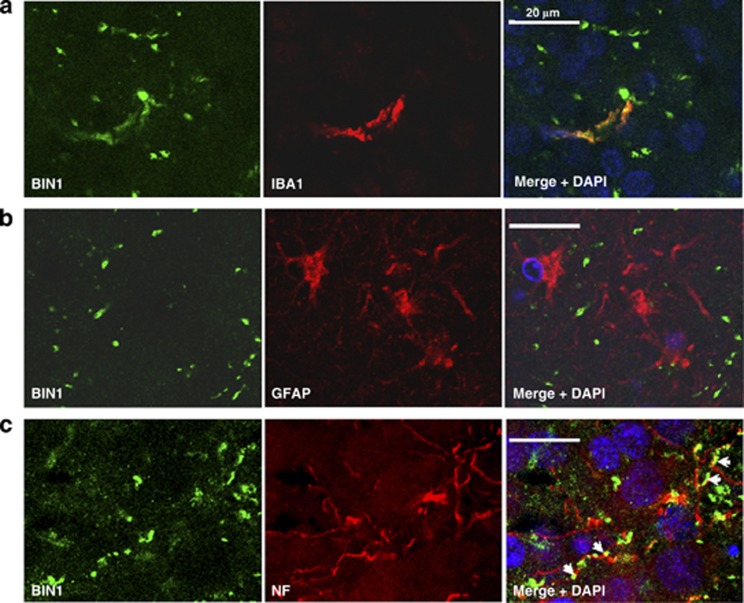
Figure 5.
Representative confoncal images of BIN1 staining in AD brains. Representative confoncal images of BIN1 staining in hippocampus from AD patients (Braak VI). (a) Anti-IBA1 and (b) anti-GFAP antibodies have been used as microglia and astrocytes markers, respectively. (c) Representative confocal images of BIN1 staining in AD brains. BIN1 staining followed the neurofilament (NF) labeling in axons (arrows).

