
Figure 2. Substrate Binding Site of DhbE.
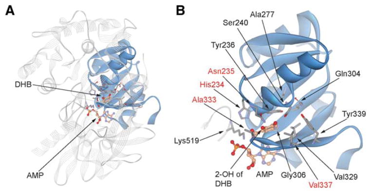
(A) Crystal structure of DhbE with the binding pocket of DHB shown in solid ribbons and the rest of the protein in line ribbons (Protein Data Bank ID 1MD8) (May et al., 2002).
(B) Detailed structure of DHB binding site showing key active-site residues as the nonribosomal code for DHB recognition. Names of the residues randomized in the DhbE library are in red.
See also Figure S4.