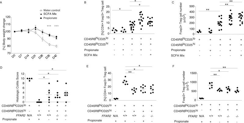
Fig. 4. SCFA exposure ameliorates T cell transfer colitis in a Treg-intrinsic, Ffar2-dependent manner.
BALB/c Rag2−/− mice were injected with CD4+CD45RBhiCD25lo naive T cells alone or in combination with Tregs. Following injection, mice received propionate, SCFA mix, or pH and sodium-matched drinking water. (A) Weekly percentage body weight change is shown across the experimental groups from experimental d0-d42. Symbols show the mean and error bars the SD. Data reflect three independent experiments. Colonic LP lymphocytes were isolated and stained for CD4 and Foxp3 and (B) percentage and (C) number of CD4+Foxp3+ within the CD45+CD3+ population are shown. Symbols represent data from individual mice, horizontal lines show the mean and error bars the SD. (D-F) C57BL/6 Rag2−/− mice were injected with CD4+CD45RBhiCD25lo naive T cells alone or in combination with Ffar2+/+ or Ffar2−/− Tregs. Following injection mice received propionate or pH and sodium-matched drinking water. (D) Histologic colitis score is shown along the y-axis, the treatment group and experimental conditions are shown along the x-axis. Colonic LP lymphocytes were isolated and (E) percentage and (F) number of CD4+Foxp3+ within the CD45+CD3+ population are shown. Symbols represent data from individual mice. Horizontal lines show the mean and error bars the SD. Panels D-F represent data from 2 independent experiments. The Kruskal-Wallis test with a Dunn's post hoc test was performed for panels A-F. ** P <0.01, * P <0.05.

