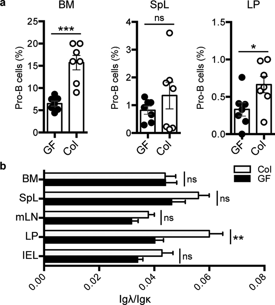
Figure 4. Effects of Gut colonization on Development of LP B Lineage Cells.
a, Plots of percentage of pro-B cells versus total CD19+ B lineage cells from bone marrow (BM), spleen (SpL) and lamina propria (LP) of 4 wk-old germfree (GF) mice and littermates colonized (Col) by co-housing with serum pathogen free mice for 7 days. b, Bar graphs show ratios of Igλ+ versus Igκ+ B cells within mesenteric lymph nodes (mLN), inter-epithelial lymphocyes (IEL) and tissues indicated in (a) from GF and Col mice. Mean values and s.e.m are shown. The P values are indicated as: *P ≤ 0.05, **P ≤ 0.01, ***P ≤ 0.001. ns = not significant. (Details in Supplementary Fig. 16 and 17)

