
Fig 6.
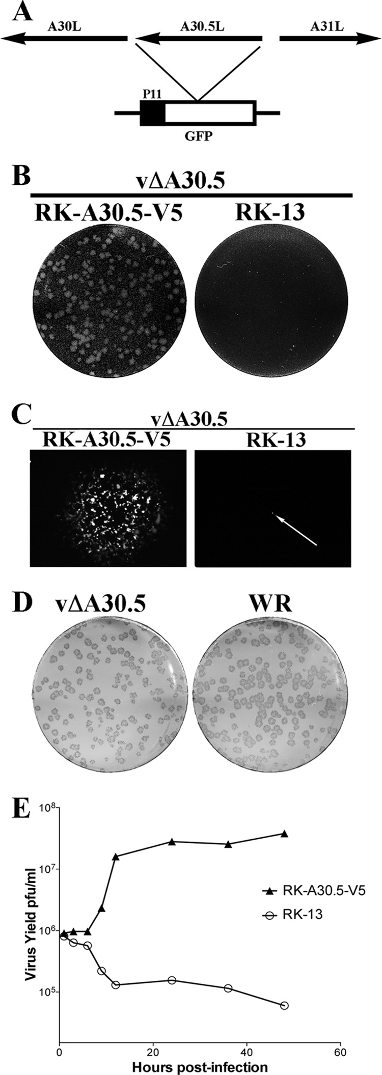
Construction and characterization of vΔA30.5. (A) Schematic representation of the genome structure of vΔA30.5. The A30.5L ORF was replaced with the GFP ORF regulated by the VACV P11 late promoter. Arrows indicate the direction of transcription. (B) Plaque phenotype of vΔA30.5. RK-A30.5-V5 and RK-13 cells were infected with vΔA30.5. After 48 h, the cells were stained with crystal violet. (C) Plaques formed by vΔA30.5 in RK-A30.5-V5 cells and a single infected cell (arrow) in RK-13 cells visualized with a fluorescence microscope. (D) Comparison of plaques formed by vΔA30.5 and wild-type VACV strain WR on RK-A30.5-V5 cells. After 72 h, plaques were visualized by staining with antibody to VACV followed by anti-protein A conjugated to alkaline phosphatase. (E) One-step growth curve of vΔA30.5. RK-A30.5-V5 and RK-13 cells were infected with 3 PFU/cell of vΔA30.5. At the indicated times, the infected cells were harvested and virus titers were determined in duplicate by plaque assay on RK-A30.5-V5 cells. Data from two independent experiments were averaged.