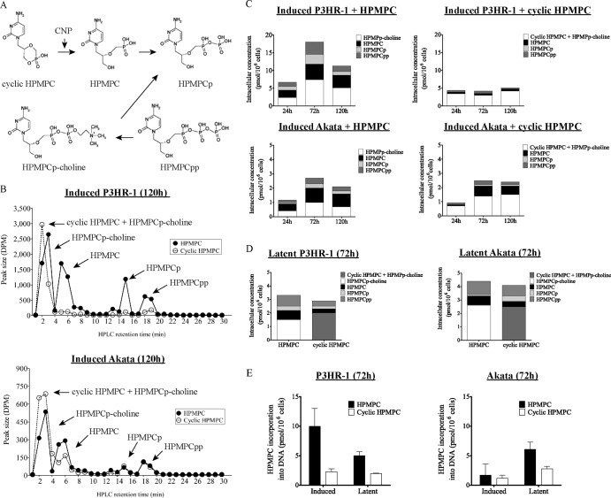
Fig 2.
Metabolism of [5-3H]HPMPC and cyclic [5-3H]HPMPC in P3HR-1 and Akata cells after 120 h. (A) Activation pathway for HPMPC and cyclic HPMPC. CNP, cellular 2′,3′-cyclic nucleotide 3′-phosphodiesterase. (B) Representative HPLC result showing the metabolism of 10 μM [5-3H]HPMPC and cyclic [5-3H]HPMPC in induced P3HR-1 and Akata cells after 120 h of exposure to the indicated compound. The arrows point to the different metabolites of HPMPC and cyclic HPMPC. Note the differences between values on the y axis. (C) At the designated time points, intracellular concentrations of HPMPC, HPMPCp, HPMPCpp, HPMPp-choline, and a mixture of cyclic HPMPC and HPMPCp-choline were determined in P3HR-1 and Akata cells after EBV reactivation. The means of two independent experiments were plotted. (D) Intracellular concentrations of HPMPC metabolites in latently infected cells at 72 h after addition of HPMPC or cyclic HPMPC were plotted as the means of two independent experiments. (E) Incorporation of HPMPC into DNA of P3HR-1 and Akata cells in the latent and lytic stages of the virus life cycle after 72 h of incubation.

