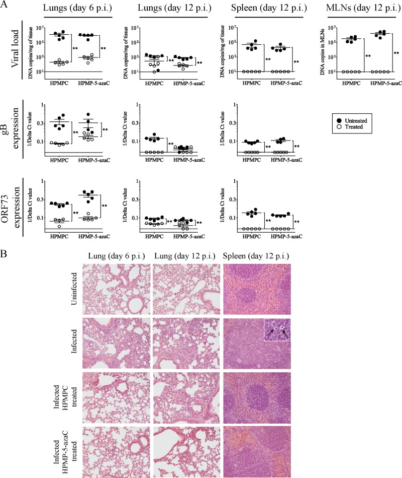
Fig 5.
Analysis of MHV-68 infection in different organs of untreated and treated mice at acute and latent stages of infection. (A) DNA copy numbers were detected in the lungs, MLNs, and spleens of mice infected by the intranasal route with 104 PFU MHV-68 and treated with HPMPC or HPMP-5-azaC (i.p.) for 5 consecutive days. Each group contained five mice. Results are presented as the mean log viral copy number per mg of tissue ± the standard deviation. The levels of MHV-68 gB and ORF73 expression, normalized to the endogenous control, glyceraldehyde-3-phosphate dehydrogenase, were measured relative to the infected control by using the comparative threshold cycle (CT) method (ΔΔCT method). Statistical significance was calculated using the Mann-Whitney U test. *, P < 0.05; **, P < 0.01. (B) Hematoxylin and eosin staining of lung tissues of uninfected mice, MHV-68-infected mice, HPMPC-treated mice, and HPMP-5-azaC-treated mice at days 6 and 12 p.i. Histopathology of the spleen at day 12 p.i. is shown in the panels on the right, and arrows indicate the presence of prominent macrophages in the spleens of infected mice. Magnification, ×20 (inset, ×100).

