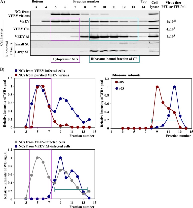
Fig 7.
Deletion of SD1 in wt capsid protein has a deleterious effect on the accumulation of NCs in the infected cells. (A) Results of one of the reproducible analyses of NC and capsid protein distribution in sucrose gradients. Cells were infected and lysed as described in Materials and Methods. The details of ultracentrifugation, fractionation of the gradients, and analysis of fractions by Western blotting were performed according to the protocols presented in detail in Materials and Methods. Titers of the released viruses were determined at the time of cell harvesting, at 16 h postinfection. The same membranes were stained with antibodies specific to ribosomal proteins, and two representative blots are presented. (B) Results of quantitative analysis of capsid and ribosomal protein distribution in various fractions in panel A. In each gradient, values were normalized to the signal detected in the fraction with the highest concentration of capsid protein. Pink boxes indicate the distribution of cytoplasmic NCs. Green lines indicate positions of the ribosome-bound fraction of capsid proteins.

