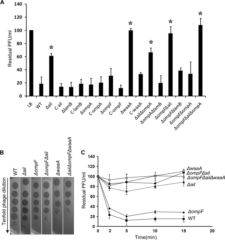
Fig 3.
(A) Yep-phi adsorption to Y. pestis outer membrane protein and lipopolysaccharide mutants, shown as residual PFU percentages. The control and strains used for adsorptions are presented below the columns (complete strain names and descriptions can be found in Table 1). Error bars denote ranges. Significance was determined by one Student t test for comparison between the mutant group and the WT group; *, P < 0.05. (B) Tenfold dilution of lysates of Yep-phi applied to bacterial lawns of wild-type and mutant Y. pestis strains. (C) Adsorption kinetics of Yep-phi to Y. pestis LPS and OMP mutants, shown as residual PFU percentages. Standard errors are indicated by vertical lines.

