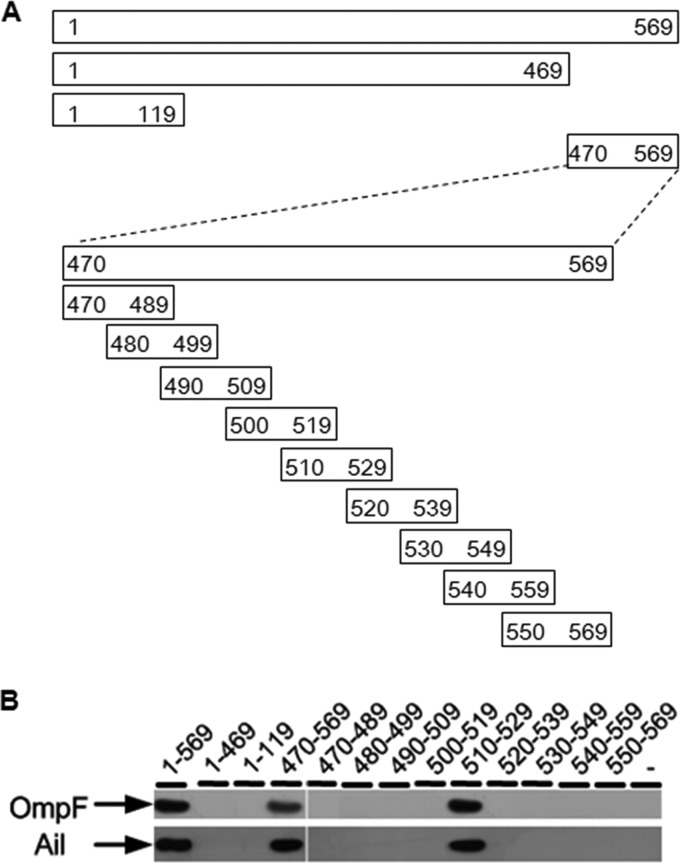
Fig 4.
(A) Positions of the fragments on the gp17 protein. The thick line represents the fragments, and the numbers on the two ends of each fragment denote the starting and ending amino acid positions. (B) OmpF and Ail detected by Western blotting using either rabbit anti-OmpF or anti-Ail polyclonal antibody after affinity chromatography.

