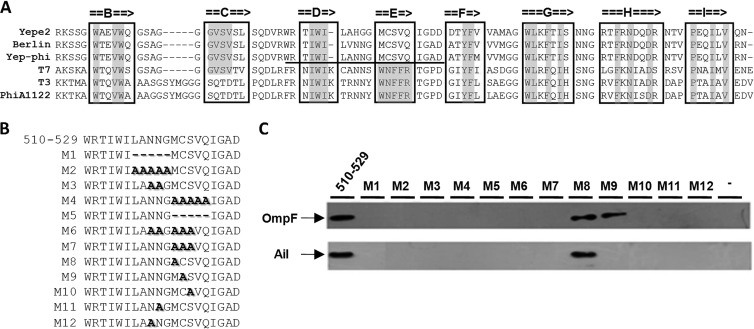
Fig 5.
(A) Alignment of the sequence of the tip domain of T7 gp17 with its homologs from E. coli phage T3 and Y. pestis phages Yep-phi, Berlin, Yepe2, and PhiA1122. Beta-strands are denoted by arrows. The residues of beta-strands that are identical with each other are marked with gray. The domain necessary for the phage Yep-phi and Y. pestis interaction is underlined. (B) Locations of deletions and alanine replacements used in this study are indicated by M1 to M12. (C) OmpF and Ail were detected by Western blotting using either rabbit anti-OmpF or anti-Ail polyclonal antibody after affinity chromatography.

