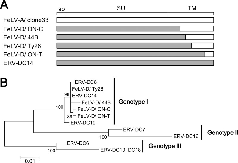
Fig 1.
Structure and phylogenetic analysis of the env gene from FeLV-D and ERV-DC. (A) Schematic representation of the env structure of FeLV-D and ERV-DC. The recombination pattern of each env is indicated. White indicates FeLV-A sequences, and gray indicates ERV-DC sequences. sp, signal peptide; SU, surface unit; TM, transmembrane. (B) Best-maximum-likelihood tree from phylogenetic analysis of env nucleotide sequences corresponding to the signal peptides and surface units from FeLV-D and ERV-DC strains.

