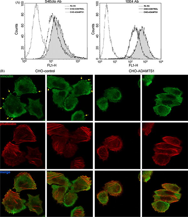
Fig. 6.

Analysis of cell surface syndecan-4 and focal adhesions in CHO-K1 cells. (A) Parental and ADAMTS1 CHO-K1 cells were labeled with S4Ecto and 10E4 antibodies and fluorescence-intensity was analyzed by flow cytometry. (B) Same cells as A were plated in fibronectin-coated coverslips and fixed after 2 h. Focal adhesions (yellow arrows) and stress fibers were visualized by staining with a vinculin antibody and Phalloidin-TRITC, respectively. (For interpretation of the references to color in this figure legend, the reader is referred to the web version of the article.)
