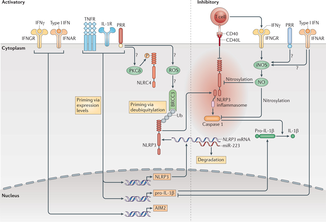
Figure 2. Extracellular signals regulate the inflammasomes.
Pro-interleukin-1β (pro-IL-1β) and NLRP3 (NOD-, LRR-and pyrin domain-containing 3) expression are induced by transcriptionally active pattern recognition receptors (PRRs) or by cytokine receptors. Furthermore, NLRP3 deubiquitylation by the K63-specific deubiquitinase BRCC3 is c rucial for its activation. Direct contact with mature or memory T cells inhibits the inflammasomes, probably via tumour necrosis factor receptor (TNFR) superfamily interactions. Type I interferons (IFNs) inhibit the transcription of pro-IL-1β , but also upregulate the expression of absent in melanoma 2 (AIM2). Both type I IFNs and IFNγ inhibit NLRP3 through the induction of nitric oxide (NO) via inducible nitric oxide synthase (iNOS), possibly with the requirement of concomitant priming by PRRs. Ub shows ubiquitylated proteins. Question marks show pathways that are still speculative. CD40L, CD40 ligand; IFNAR, interferon-α/β receptor; IFNGR, interferon-γ receptor; IL-1R, IL-1 receptor; miR-223, microRNA-223; NLRC4, NOD-, LRR- and CARD-containing 4; ROS, reactive oxygen species; PKCδ, protein kinase Cδ.

