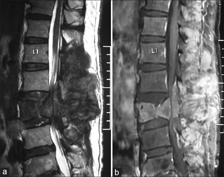
Figure 5.

Magnetic resonance imaging of spine in sagittal view shows a T1 (b) hypo, T2 hyper (a) heterogenously enhancing lumbar (L1- L4) extradural mass lesion with associated L3 vertebral body compression collapse

Magnetic resonance imaging of spine in sagittal view shows a T1 (b) hypo, T2 hyper (a) heterogenously enhancing lumbar (L1- L4) extradural mass lesion with associated L3 vertebral body compression collapse