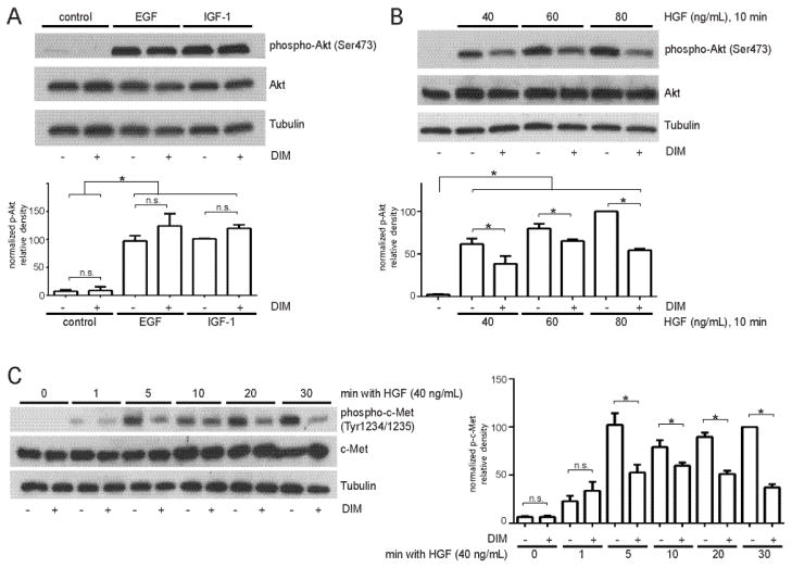
Fig. 2.
DIM inhibits Akt activation by HGF and decreases phosphorylation of c-Met. (A) MDA-MB-231 cells were incubated overnight in serum-free medium, treated with DIM for 4 h, and then epidermal growth factor (EGF, 25 ng/mL) or insulin-like growth factor-1 (IGF-1, 25 ng/mL) for 10 min. (B) Cells were incubated overnight in serum-free medium, treated with DIM for 4 h, and HGF (40 ng/mL) for 10 minutes. Total cell lysates were collected and analyzed by Western blotting for phospho-Akt Ser473 and total Akt. (C) Western blot analysis of phospho-c-Met Tyr1234/1235 and total c-Met from cells incubated in serum-free medium overnight and then treated with DMSO or 25 μM DIM for 4 h, followed by HGF treatment for 0–30 min. Tubulin was used as a loading control. Histograms depict phospho-Akt or phospho-c-Met band density normalized to total Akt or total c-Met band density expressed as percent of control. Bars represent mean ± SEM of 3 independent experiments. *, p<0.05; n.s., not statistically significant.

