Figure 1.
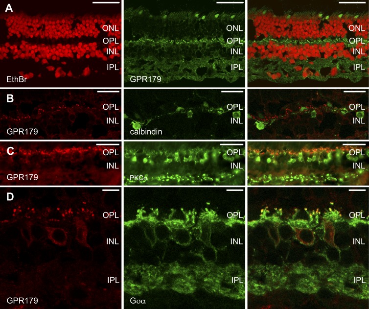
GPR179 colocalizes with markers for ON-BCs but not for HCs. Left and middle columns show the individual channels while the right column shows the combination of the 2 channels. (A) Immunohistochemical localization of GPR179 (green) in the human retina. Nuclei (red) were stained with ethidium bromide. Note the punctate labeling in the OPL and diffuse, nonspecific labeling in the IPL. (B) Retinal section labeled with antibodies against GPR179 (red) and calbindin (green). Calbindin labels some HC types. No calbindin labeling could be detected in ON-BCs. Calbindin-IR does not overlap with GPR179-IR, suggesting that GPR179 is not expressed by HCs or OFF-BCs. (C) Retinal section labeled with antibodies to GPR179 (red) and PKCα (green). PKCα labels rod-BCs and colocalization is seen at the tips of the ON-BC dendrites. (D) Retinal section labeled with antibodies to GPR179 (red) and Goα (green). Goα labels ON-BCs and colocalization was found at the tips of the ON-BC dendrites. Scale bars: 50 μm (A, C), 15 μm (B), 5 μm (D). ONL, outer nuclear layer; INL, inner nuclear layer.

