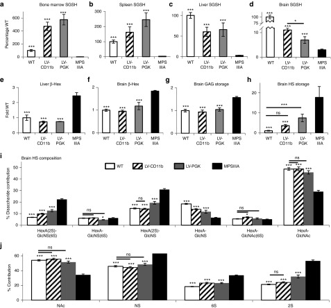
Figure 3.
LV-CD11b improves brain-specific SGSH activity over LV-PGK and corrects primary HS storage of MPSIIIA mice. At 8 months of age, SGSH enzyme activity was measured in (a) bone marrow, (b) spleen, (c) liver, and (d) brain of perfused mice (n = 5). Secondary upregulation of β-Hex was measured in (e) liver and (f) brain. (g) Total sulfated GAG in the brain was measured with the dye binding Blyscan assay. (h) Total HS storage was measured by 2-aminoacridone (AMAC)–labeled disaccharide analysis. The different HS disaccharides were quantified by (i) AMAC, and (j) the relative proportion of HS that was NAc, NS, 6S, and 2S was also determined. Error bars represent the SEM. Significant differences to MPSIIIA are marked with *P < 0.05 and ***P < 0.001. Treatments that are not significantly different to WT are marked with a line and ns (nonsignificant) (P > 0.05). 6S, 6-O-sulfated; 2S, 2-O-sulfated; β-Hex, β-N-acetyl hexosaminidase; GAG, glycosaminoglycan; HS, heparan sulfate; LV, lentiviral vector; MPSIIIA, mucopolysaccharidosis type IIIA; NAc, N-acetylated; NS, N-sulfated; PGK, phosphoglycerate kinase; SGSH, N-sulfoglucosamine sulfohydrolase; WT, wild-type.

