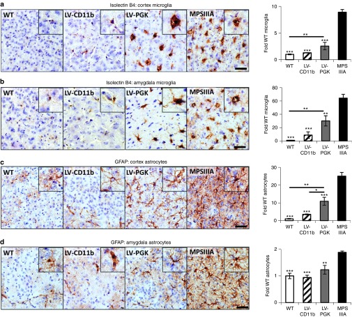
Figure 5.
LV-CD11b normalizes neuroinflammation in the cerebral cortex and amygdala of MPSIIIA mice, while LV-PGK mediates improvements in neuroinflammation. Four brain sections per mouse (n = 5 per group), taken from approximately bregma 0.26, −0.46, −1.18, and −1.94 mm, were stained and quantified with isolectin B4 for microglia number in the (a) cerebral cortex and (b) amygdala or with GFAP for astrocytes in the (c) cerebral cortex and (d) amygdala. Representative images from the cerebral cortex (layer IV/V) and the amygdala, both from approximately −1.18 mm relative to bregma, are shown. Sections were counterstained with Mayer's hematoxylin to highlight the nuclei (blue). Error bars represent the SEM. Significant differences to MPSIIIA, between the two groups marked with a line, are demonstrated with *P < 0.05, **P < 0.01; and ***P < 0.001. Bar = 50 µm. LV, lentiviral vector; MPSIIIA, mucopolysaccharidosis type IIIA; PGK, phosphoglycerate kinase; WT, wild-type.

