
Figure 3. Mkrn3 Messenger RNA (mRNA) Levels in the Hypothalamic Arcuate Nucleus of Male and Female Mice during Postnatal Development.
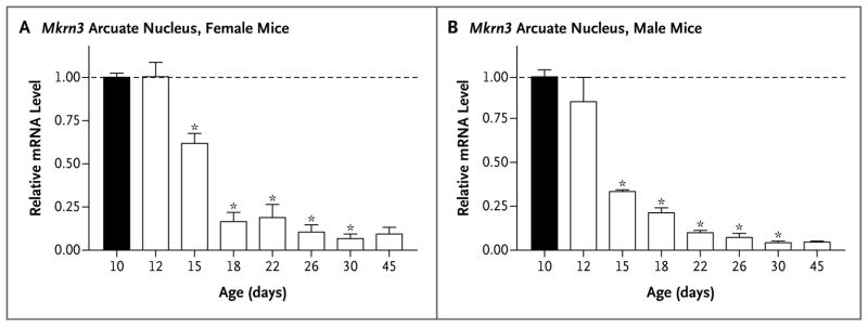
Total RNA was extracted from the hypothalamic arcuate nucleus of male and female mice at the ages indicated (number of days after birth), and Mkrn3 mRNA was quantified with the use of real-time polymerase-chain-reaction assay. The bar graphs show the relative change in mRNA levels in female mice and male mice, as compared with the level on postnatal day 10 (black bars), normalized to levels of endogenous ribosomal protein L19 mRNA. Mean (±SE) values are shown for three different mice at each age, with each measurement performed in triplicate. Significant differences (P<0.05) were measured by means of a one-way analysis of variance with a post hoc Tukey multiple-comparison test. Asterisks indicate P<0.001.