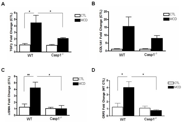
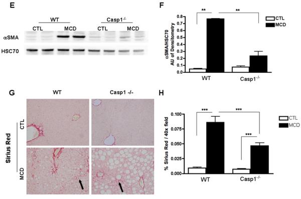
Fig. 3. HSC activation and collagen deposition during diet-induced steatohepatitis are markedly decreased in Casp1−/−.


(A-D) RT-PCR analysis of HSC activation markers transforming growth factor beta (TGFβ), collagen type I alpha 1 (COL1A1), alpha smooth muscle actin (αSMA), cysteine- and glycine-rich protein 2 (CRP2), mRNA expression in liver of Casp1−/− mice compared to WT mice (n = 5-7 in each group). (E) Western blot of αSMA on whole liver lysates in WT and Casp1−/− MCD-fed mice compared to CTL-fed mice and (F) corresponding densitometric analysis to HSC70 (n=3). (G) Collagen fibers were stained with Sirius red and (H) quantified using the surface area stained per 40x field area (n = 5-7 in each group) excluding blood vessels. Results are expressed as mean ± S.E.M. * P < 0.05; ** P < 0.01; ***P < 0.001 compared to controls.
