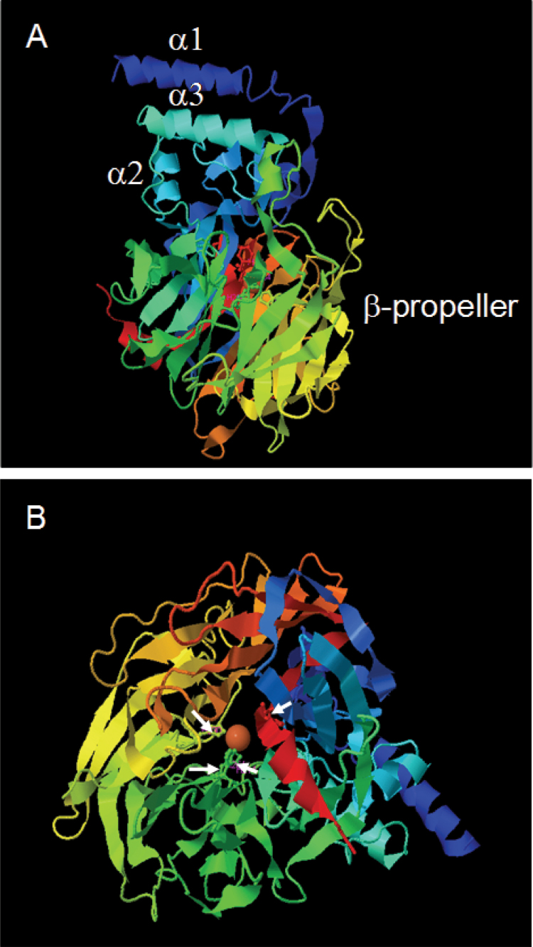
Fig. 6.
Best 3D model of CCD4b1 using the VP14 (PBD: 2biwA) structure from maize as template. The α-helix (α1, α2, and α3) and β-propeller domains are shown in model view (A). A top view of the model allows the visualization of the propeller blades and the Fe2+ in the reaction centre (B). White arrows in (B) indicate the location of the four histidines coordinating the Fe2+. (This figure is available in colour at JXB online.)

