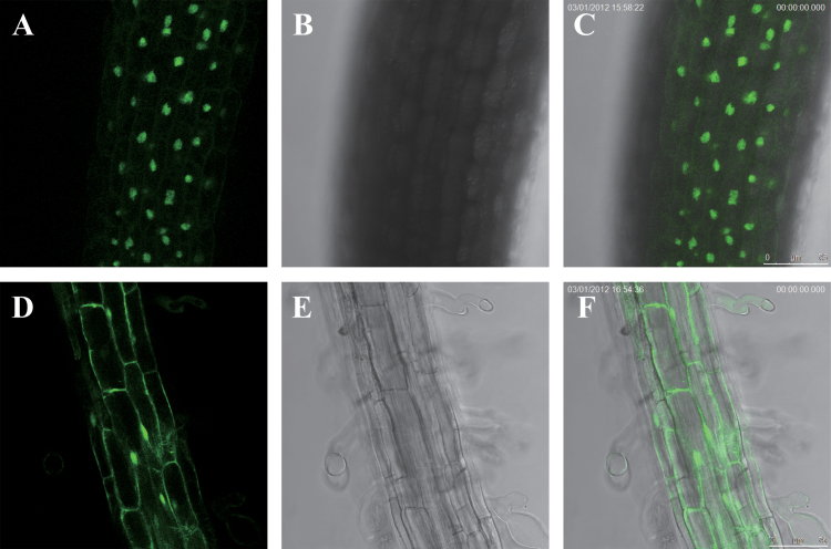
Fig. 4.
The subcellular localization of 35S:PdNF-YB7-GFP and control 35S:GFP expression in Arabidopsis cells. Cells were detected under a fluorescent or light field by a fluorescence microscope. (A–C) 35S:PdNF-YB7-GFP seedling in fluorescent light (A), bright light (B), and overlap image of fluorescent and bright light (C). (D–F) Control seedling in fluorescent light (D), bright light (E), and overlap image of fluorescent and bright light (F). Bars, 75 μm (this figure is available in colour at JXB online).

