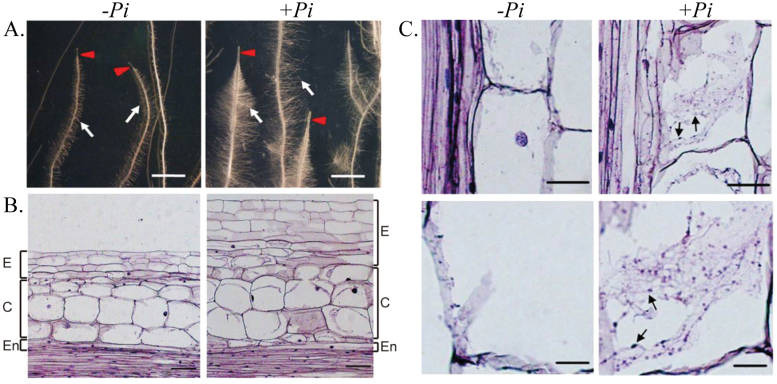
Fig. 4.
Root architecture and anatomy of Chinese cabbage colonized by Piriformspora indica at 7 d post-inoculation. (A) Morphology of cabbage root colonized with (+Pi) or without (–Pi) P. indica. Left panel: control root (–Pi) showing a poorly developed root tip (red arrow), formation of unbranched lateral roots, and less root mass (white arrow). Right panel: P. indica-colonized root (+Pi) showing a long and enlarged root tip (red arrow), fully developed branches of lateral root formation, and dense root biomass (white arrow). Scale bar = 2mm. (B) Longitudinal section of Chinese cabbage root stained with aniline blue. Lateral root section illustrating its structural differences with or without colonization by P. indica: epidermis (E), cortex (C), and endodermis (En). Left panel: control root (–Pi) showing normal characteristics of epidermal cells, cortex, and endodermis. Right panel: P. indica-colonized root (+Pi) showing visible expansion of the epidermis and enlargement of the cortex surface area. Scale bars = 10 μm. (C) Microscopic observation of P. indica-colonized root stained with aniline blue. A root longitudinal section showing hyphal penetration inside root cells and cell enlargement/expansion induced by P. indica colonization. Root tissues (n=40) used for microdissection were taken 14 d post-inoculation by P. indica. Left panel: control root (–Pi) cell showing no hypha and spores. Right panel: P. indica-colonized root (+Pi) cells of the cortex and epidermis showing intracellular hyphae and spores (arrow). Scale bars = 10 μm.

