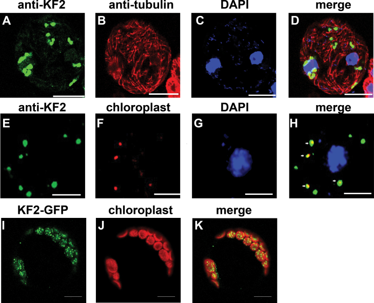
Fig. 10.
Subcellular localization of CsKF2 in plant cells. (A–D) Triple immunolocalization of CsKF2, tubulin, and the nucleus in fruit cells at 0 DAA. Anti-KF2 antibodies detected particles in a punctate pattern in the cytoplasm in fruit cells at 0 DAA (A; green). Microtubules were immunofluorescently labelled with anti-α-tubulin antibody and TRITC-conjugated goat anti-mouse IgG (B; red). The nucleus was detected by DAPI (C; blue). No CsKF2 signal was associated with microtubules (D; merge). (E–H) Co-localization of CsKF2 with chloroplast in fruit cells at 0 DAA. Anti-KF2 antibodies detected particles in a punctate pattern in the cytoplasm in fruit cells at 0 DAA (E; green). Chloroplast autofluorescence was viewed as the red channel (F; red). The nucleus was detected by DAPI (G; blue). CsKF2 green signals showed co-localization with chloroplasts (H; merge). (I–K) Co-localization of CsKF2–GFP with the chloroplast in Arabidopsis protoplasts. The protoplasts were transiently transformed with the CsKF2–GFP construct (I; green), and the chloroplast autofluorescence was viewed through the red channel (J; red). Strong CsKF2 signals appeared in particles in the chloroplasts (K). Scale bar=5 μm.

