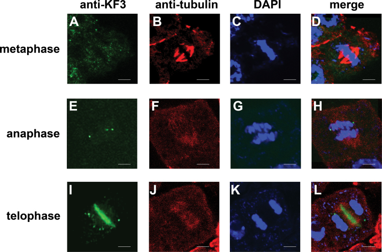
Fig. 12.
CsKF3 distributes in a cell cycle-dependent manner in cucumber fruit cells. CsKF1 (green), microtubules (red), and the nucleus (stained with DAPI; blue) are shown at different cell division stages. (A–D) In metaphase cells, few CsKF3 punctate signals could be detected by fluorescence microscopy. (E–H) During anaphase, only small CsKF3 punctate signals could be detected by anti-CsKF3 antibodies. (I–L) CsKF3 mainly localized at the midzone of the phragmoplast in telophase cells. Scale bar=5 μm.

