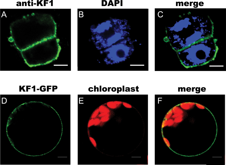
Fig. 9.
Subcellular localization of CsKF1 in plant cells. (A–C) Immunolocalization of CsKF1 in fruit cells at 5 DAA. The 9930 fruit cells harvested at 5 DAA were stained with the anti-CsKF1 antibody (A; green) and DAPI (B; blue). Strong CsKF1 signals appeared close to the plasma membrane (C). (D–F) In living Arabidopsis protoplasts, CsKF1–GFP localized to the plasma membrane. The protoplasts were transiently transformed with the CsKF1–GFP construct (D; green), and the chloroplast autofluorescence was viewed as the red channel (E; red). Strong CsKF1 green signals localized in the plasma membrane (F). Scale bar=5 μm.

