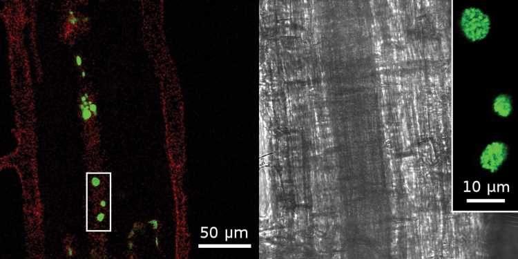
Fig. 2.
Green fluorescent spots in M. sinensis roots inoculated with GFP-labelled H. frisingense. Bacterial aggregates were visible in the intercellular apoplastic space. Bright field image (right) and fluorescence image (left). The red colour indicates residual background fluorescence from the cell wall. The inset shows a magnification of the bacterial colony aggregates. (This figure is available in colour at JXB online.)

