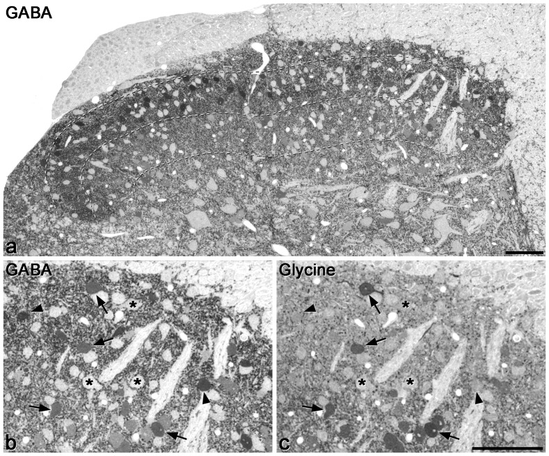
Figure 1. GABA and glycine immunoreactivity in a 0.5 µm thick transverse section of the mouse dorsal horn.
a shows the whole mediolateral extent of the dorsal horn stained for GABA, while b and c show the medial part of the dorsal horn at higher magnification, in the same section and in a section reacted for glycine, respectively. Many GABA-immunoreactive cell bodies are scattered throughout the dorsal horn, and although these vary in intensity, they are clearly darker than the immunonegative cells, some of which are marked with asterisks in b. The dark staining in the neuropil represents GABAergic axons and dendrites. Dashed lines show the ventral borders of laminae I, II and III. b and c are serial semithin sections, so the same cells are visible. A few cells with relatively strong glycine immunoreactivity are visible (some marked with arrows) and these are also GABA-immunoreactive. In addition, there are cells that are GABA- but not glycine-immunoreactive (two marked with arrowheads). Scale bars = 100 µm.

