
Figure 5. Restriction of sst2A-immunoreactivity to GABAergic neurons in the superficial dorsal horn.
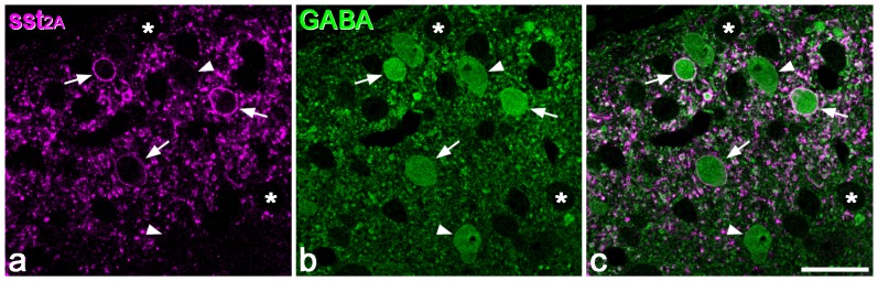
a: A confocal scan (single optical section) from the top surface of a Vibratome section that had been reacted to reveal sst2A (magenta) and GABA (green). Three sst2A-immunoreactive cell bodies are marked with arrows, and there are many other labelled profiles, most of which are dendrites cut in cross section. b: The same field scanned to reveal GABA. Several GABA-immunoreactive cell bodies can be seen and some of these are indicated with arrows or arrowheads. Many cell bodies that are not GABA-immunoreactive are also present, and two are marked with asterisks. c: The merged image reveals that the sst2A-immunoreactive cells are all immunostained for GABA, but that several GABA+ cells do not possess sst2A (two are indicated with arrowheads). Scale bar = 20 µm.