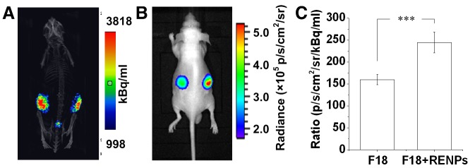
Figure 5. In vivo CLI of the subcutaneous Matrigel pseudotumor animal models.

(A) MicroPET/CT imaging of subcutaneous Matrigel pseudotumors containing 37 MBq 18F-FDG (left flank) or 18F-FDG with 2 mg/mL REMPs (right flank) in mice. (B) CLI of the same subcutaneous Matrigel pseudotumor mouse model. (C) Quantitative analysis of the Cerenkov luminescence intensity of the pseudotumors on both sides. The pseudotumor of 18F-FDG with REMPs showed stronger luminescence intensity than that of 18F-FDG with DMSO (***P < 0.001).
