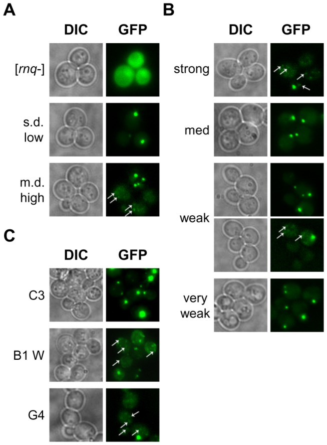
Figure 3. Spontaneously formed [RNQ+] variants show diversity in Rnq1-GFP aggregation patterns.

Cells were transformed with a plasmid expressing Rnq1(153-405)-GFP under the CUP1 promoter. Prior to imaging, transformants were transferred to induction medium containing 50μM CuSO4 and grown for ~2.5 hours. A variety of fluorescence patterns were observed, including: a single focus, large multiple foci, and numerous petite foci. Arrows identify petite foci that are fainter and smaller. Representative images in differential interference contrast mode (DIC) and under a GFP-emitted light filter (GFP) are shown for (A) previously published [RNQ+] variants and [rnq-], (B) all of the stable [RNQ+] variants, and (C) a subset of the unstable [RNQ+] variants.
