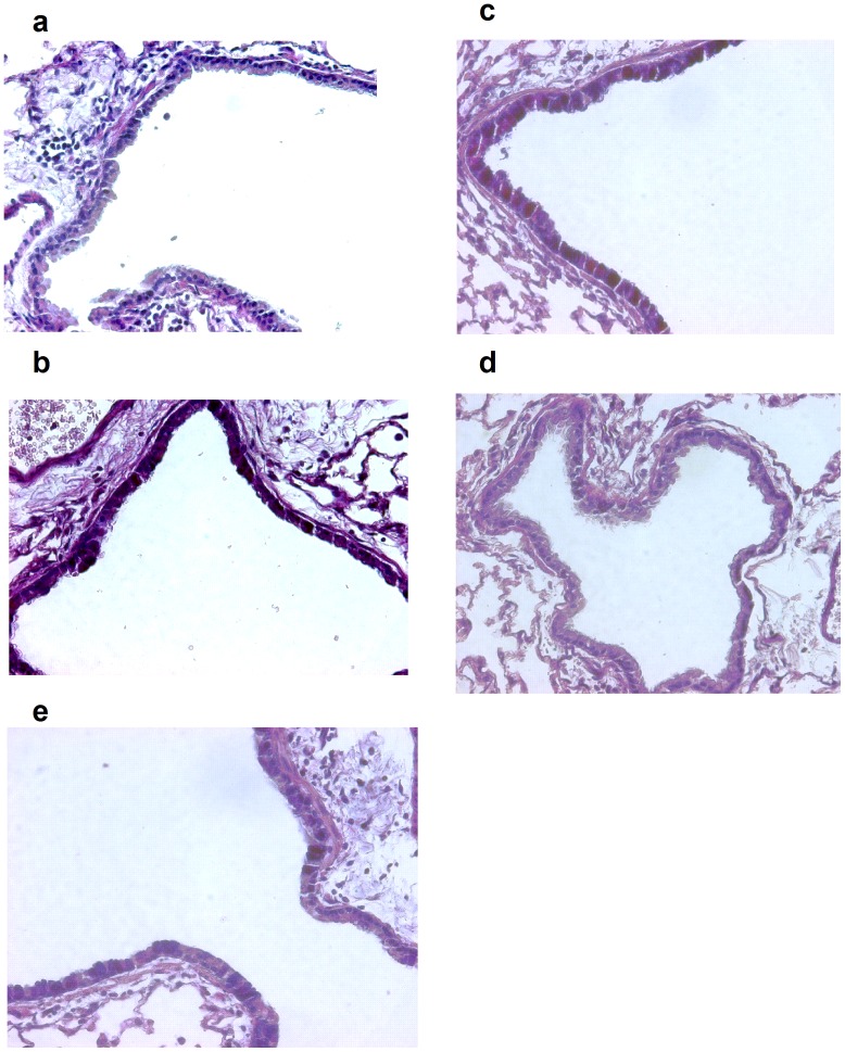
Figure 9. PAS staining for goblet cells from representative sections of lobar bronchi or daughter generation airway in mice from a) air-exposed, b) Ova-exposed (Ova), c) Ova-exposed Dex-treated (Dex), d) Ova-exposed Dex-nanoparticle (Dex NP), and e) Ova-exposed nanoparticle (NP) treated mice.
Lung sections were stained with Alcian Blue-Periodic Acid-Schiff (PAS) and counterstained with hematoxylin and eosin and photographed at 400×magnification. PAS positive cells in airways were fewer in Dex NP treated animals.

